
Comparing Central Corneal Thickness Using Ultrasound And Anterior Segment Optical Coherence Tomography Pachymetry In Adults Attending A Private Eye Clinic In Abuja. https://doi.org/10.60787/NMJ-63-1-83
Main Article Content
Keywords
Central Corneal Thickness, Ultrasound Pachymetry, Optical Coherent Tomography
Abstract
Background: Central Corneal Thickness (CCT) measurement is useful in managing glaucoma, ocular hypertension, corneal lesions and kerato-refractive surgeries. The study aims to compare the CCT measurements between Ultrasound Sonography (USS) and Optical Coherence Tomography (OCT) to analyze the correlation and agreement between these instruments and the repeatability of each instrument.
Methodology: A cross-sectional comparative study was carried out in 100 eyes of 50 patients attending Rachel Eye Center in Abuja from January to March 2021. The CCT was taken using the USS and OCT. CCT was measured using the Pachscan ultrasound and the Optovue machine utilised for the OCT technique. Data were analysed using SPSS version 20 using Paired Sample t-Test, Pearson’s correlation, Interclass Correlation and Bland Altman Methods.
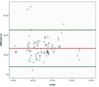
Results: Patients were between 18 and 79 (mean age of 39.1), 72 males and 28 females. The mean CCT was 537.36 ±33.26 and 510.94 ±33.13 for USS and OCT, respectively, with a mean difference of (26.42±9.53 p<0.001). There was a very strong correlation between the two sets of measurements, r = 0.997 p<0.001. There was a high average mean intraclass correlation coefficient of 0.843 between the two instruments, and this was excellent (0.961) within the 95 percentile upper limit but poor in the (0.096) lower limit. There was a high correlation and no statistically significant difference in comparing repeated mean USS and OCT measurements. There was an excellent average mean Intraclass Correlation Coefficient of 0.999/0.997 for the repeated OCT and USS values, which was found to be excellent(0.999/0.998) within the 95 percentile upper limit and (0.997/0.994)lower limit, respectively.
Conclusion: We find that measurements of CCT using the Pachscan ultrasound and Optovue OCT correlate well, but the mean Pachscan measures were significantly higher than Optovue measures.
References
2.Gillis A, Zeyen T. Comparison of optical coherencereflectometryandultrasoundcentral corneal pachymetry. Bull Soc Belge Ophthalmol 2004; 292:71-5.
3.SebileÜstünÇomçalý, RaþitKýlýç, SerdarBayraktar, YasinÇakmak, ZelihaYazar, HafizeSezer, AyhanDursun “Comparison of UltrasonicPachymetryandOpticalCoherence Tomography for the Measurement ofCentralCornealThickness”Glo-Kat2016;11:93-96
4. Ramesh PV, Jha KN, Srikanth K. Comparison of Central Corneal Thickness using Anterior Segment Optical CoherenceTomographyVersus Ultrasound Pachymetry. J Clin Diagn Res.2017;11:NC08-NC11.doi:10.7860/JCDR/2017/25595.10420
5.NortheyLC,GiffordP,BonehamGC.ComparisonofTopconopticalcoherencetomographyandultrasoundpachymetry.Optom Vis Sci2012; 89:1708-14.
6.Babbar S, Martel MR, Martel JB (2017) Comparison of central corneal thickness by ultrasound pachymetry, optical coherence tomography and specular microscopy. New FrontOphthalmol3:DOI:10.15761/NFO.1000164
7.Garcia-Medina JJ, Garcia-Medina M, Garcia-Maturana C, et al. Comparative study of central corneal thickness using Fourier-domain opticalcoherence tomography versus ultrasound pachymetry in primary open angle glaucoma. Cornea2013;32:9-13.
8.Vollmer L, Sowka J, Pizzimenti J, et al. Central corneal thickness measurements obtained with anterior segment spectral domain optical coherence tomography compared to ultrasound pachymetry in healthy subjects. Optometry2012;83:167-72.
9.Pateras, E., & Kouroupaki, A. I. (2020). Comparison of Central Corneal Thickness Measurements between Angiovue Optical CoherenceTomography,UltrasoundPachymetryandOcularBiometry.Ophthalmology Research: An International Journal, 13, 1-9.
10.AcarBT,TuranEV,HaliliEveark.Merkezikorneakalýnlýðýölçümündeultrasonikpakimetriileoptikkoherenstomografininkarþýlaþtýrýlmasý.MN Oftalmol2011;18:142-5.
11.Khaja W, Grover S, Kelmenson A, Ferguson L, Sambhav K, Chalam K. Comparison of central corneal thickness: ultrasound pachymetry versuss lit-lampoptical coherence tomography, specular microscopy, and Orbscan. Clin Ophthalmol. 2015; 9:1065-1070
12.Keskin S, Akýl H, Yarangümeli A, ve ark. Standart ultrasonik pakimetrive optik koheren stomografii leölçülen santralkorneakalýnlýklarýnýnkarþýlaþtýrýlmasý. MN Oftalmol 2013;20:1-3.
13.Leung DY, Lam DK, Yeung BY, et al. Comparison between central corneal thickness measurements by ultrasound pachymetry and optical coherence tomography. Clin Experiment Ophthalmol2006; 34:751-4.
14.Ayala M, StrandåsR .Accuracy of optical coherence tomography (OCT) in pachymetry for glaucoma patients BMC Ophthalmology2015;15:124 .
15.Oluwadiya K Getting to know SPSS Astep by step guide 2nd Edition.2010 ISBN:978-302-6744
16.Bland JM, Altman DG. Statistical methods for assessing agreement between two methods of clinical measurement. Lancet 1986;1:307-10.
17.Koo TK, Li MY. AGuideline of Selecting and Reporting Intraclass Correlation Coefficients for Reliability Research [published correction appears in J Chiropr Med. 2017 Dec;16:346]. J Chiropr Med.2016;15:155-163.
18.Lin C-W, Wang T-H, Huang Y-H, Huang J-Y. Agreement and repeatability of central corneal thickness measurements made by ultrasound pachymetry and anterior segment optical coherence tomography. Taiwan J Ophthalmol.2013;3:98–102
Article Sidebar
Article Details

This work is licensed under a Creative Commons Attribution-NonCommercial-ShareAlike 4.0 International License.
This is an open-access journal and articles are distributed under the terms of the Creative Commons Attribution Non-Commercial Share-Alike License 4.0. This licence allows users to download and share, remix, tweak and build upon the article for non-commercial purposes, so long as the original authorship is acknowledged and the new creations are licensed under identical terms.
Adaora Chinwendu Okudo, Rachel Eye Center, Abuja.
Dr Adaora Chinwendu Okudo
Consultant Ophthalmologist
Rachel Eye center, Abuja.
Olufemi Emmanuel Babalola, Rachel Eye Center, Abuja.
Prof Olufemi Emmanuel Babalola
Chief Medical Director
Rachel Eye Center, Abuja
Professor of Ophthalmology,
Department of surgery, Bingham University, New Karu/Jos, Nigeria.